Thursday, 24 September 2020

Erlotinib 150 mg medication guide for patients.

Erlotinib is a small molecule medication that inhibits the tyrosine kinase, a kind of enzyme associated with the EGFR (Human Epidermal Growth Factor Receptor).

The drug is indicated for adults and can be prescribed if you have advanced age non-small cell lung cancer. It can also be useful if your previous chemotherapy has not worked to stop your disease.

Erlotinib can also be recommended together with the other treatment named gemcitabine, if you have a cancer of pancreas at the metastatic stage. 

Here are some guidelines for patients who are taking Erlotinib:-

  • Your doctor must know about if you are pregnant or plan to become pregnant. It may be harmful to an unborn baby when taken during pregnancy. If you become pregnant use adequate contraception while on treatment, and for at least 14 days after taking the last tablet.
  • Erlotinib is known to interact with grapefruit juice so you need to avoid drinking this whilst taking this tablet. Always consume this drug exactly as your doctor has instructed you. 
  • The tablet of erlotinib should be consumed at least 1 hour prior to or 2 hours after ingestion of food. The usual dose is 1 tablet of erlotinib 150 mg every day if you have non-small cell lung cancer.
  • The usual daily dose is 1 tablet of erlotinib 100 mg, if you have metastatic pancreatic cancer. It is given together with the treatment of gemcitabine. 
  • If you take excess erlotinib than you should, contact your pharmacist immediately. You may have increased adverse reactions and your healthcare provider may interrupt your treatment.
  • Contact your doctor as soon as possible if you suffer from any side effects. In some cases your healthcare provider may need to reduce your dose of erlotinib or interrupt the treatment. 
  • The blood levels can also be impacted by the smoking of cigarettes. Be sure in order to inform your doctor if you're currently smoking, or if you quit smoking while consuming this medicine, as the dose you take may be required to be adjusted.
  • Erlotinib 150 mg is available through select specialty wholesalers or pharmacies. The erlotinib price may vary from innovator brand name to generic version. The strip of 10 tablets of Brand name TARCEVA 150 MG Tablets can be grabbed at around ₹35300.


Monday, 21 September 2020

Chronic Lymphocytic Leukemia: Causes, Symptoms, treatments and Medications

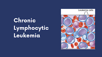
Chronic Lymphocytic Leukemia

Recent stats suggest that out of the total number of Leukemia cases, Chronic Lymphocytic Leukemia or CLL will account for a quarter of it. Most people with this condition live up to 10-20 years after getting diagnosed and getting proper treatment. However, there are some cases where disease progression is rapid, and the patients live only up to a few years. 

Causes of CLL: 

It usually starts due to a certain genetic mutation in the blood-producing cells that leads to the production of ineffective and abnormal lymphocytes. Unlike normal lymphocytes, they don’t die. Instead, they continue multiplying and starts accumulating in the blood and specific organs gradually, which causes complication. The process also interferes with the healthy cells in the bone marrow, and as a result, the production of normal blood cells gets affected. 

The risk factors may include exposure to certain chemicals like herbicides and insecticides. Having a family history of blood or bone marrow cancer may increase your chances too. 

Symptoms

  • Swollen glands in the armpits, neck, and groin
  • Unusual bruising and bleeding often
  • Night sweats
  • Shortness of breath and pale skin
  • Anaemia 
  • Unusual weight loss

These are some of the recognizable symptoms. If you face any of these, it is recommended to contact your healthcare professional and get a checkup done. 

Relation between Chronic Lymphocytic Leukemia and Myeloma 

Both the conditions—Multiple Myeloma and CLL consist of overlapping epidemiologic features. Even though they are seen to be originating from the mature B cells, the chances of these two cases coinciding are quite rare. In Multiple Myeloma, the plasma cells are affected, whereas, in CLL, the lymph nodes, liver and spleen are affected, which ultimately causes the bone marrow to stop functioning. 

Although both CLL and Myeloma are blood cancers, the latter is formed in the bone marrow and responsible for the subset of WBC to form a distinctive protein that causes cancer to grow and spread. They have parallel as well as divergent features. 

The common features include stage-dependent anaemia and immunodeficiency. Not only that, but both diseases respond to alkylating agents. 

Treatment of Chronic Lymphocytic Leukemia 

Although there are different options available, one must also have to keep in mind that there are side effects of each one of them. The upsides and downsides are measured with respect to various medical tests conducted, and then the doctor prescribes which way to go.

1. Radiation Therapy 

If the condition has been diagnosed in a localized stage and the only thing to deal with is an enlarged spleen or swollen lymph nodes, you can opt for low-dose radiation therapy. This treatment of Leukemia is safe and shows much lesser side effects. 

2. Surgery

Also referred to as Splenectomy, one may go with this option to remove the enlarged spleen. However, this is not advisable for an advanced stage condition. 

3. Stem Cell Transplant 

If it is a high-risk condition, stem cell transplant could be considered as an option for early treatment. 

4. Drug Therapy: Chemo and Targeted 

Chemotherapy and targeted therapy are two of the most commonly approached treatment options. The drugs can either be prescribed alone or in combination with other medications to yield the best results. Mention may be made of the following drugs in this respect:

Ibrutinib: It is classified as a Bruton’s Tyrosine Kinase that can inhibit the B-cell lymphoma 2 protein. It is prescribed alone or in combination with Rituximab. The typical dosage for this condition is 420mg per day, i.e., three Ibrutinib 140 mg tablets per day. However, it may be decreased depending upon the extent of the condition and response from the body. 

Alemtuzumab: It has been approved as both first-line and second-line treatment for Chronic Lymphocytic Leukemia. The drug is administered into the body as an injection in the vein. Doctors can prescribe it alone or with Rituximab. This targeted cancer drug can be classified as a monoclonal antibody. 

Venetoclax: It is a B-cell Lymphoma-2 oral inhibitor that is prescribed as a targeted drug alone or in combination with Obinutuzumab and Rituximab. High-risk older patients may also benefit from the combination of Venetoclax and Ibrutinib. The starting dose is 20mg daily for a week, which then ramps up to Venetoclax 100 mg to 400mg per day. The average treatment time is 24 months. 

Obinutuzumab: This is an antineoplastic cytotoxic drug that acts as an anti CD20 monoclonal antibody. It is a chemotherapy drug that is given to the patient as an intravenous infusion through a dedicated line. The B cells have CD20 proteins with which the drug binds to destroy them by engaging the adaptive immune system. 

These are some of the medications that have shown to slow down disease progression in CLL patients and helped to increase the survival rate over the years. Even though there are side effects to this, they are not consistent and easily manageable. 

Conclusion 

Typically, out of 10, 7 patients suffering from CLL have a survival rate of 5 years or more after getting diagnosed. The length of survival time also depends on your overall health condition, medical history and the extent of the disease. If the diagnosed patient has been in a healthy condition for most of their lives, the chances of survival are more for them. The treatment options do not guarantee a surefire cure; they are only for managing the condition and treating the symptoms so that they don’t get worse over time.  

Read:-  Venetoclax for Chronic Lymphocytic Leukemia
Thursday, 10 September 2020

Brain Metastases- All you need to know about

Brain Metastases

What is Brain Metastases: 

As any cancerous tumor grows, cancer cells can break away and be carried to the other organs of the body by the blood or lymphatic system. This is known as metastasis, or metastases when there are so many areas of spread. The metastatic brain tumors basically are the most common brain tumors. 

Although brain metastases may begin due to any kind of cancer, the types that most commonly spread to the brain are as lung, breast and kidney cancers as well as melanoma.

When any new tumor starts spreading to the brain, it isn’t addressed as brain cancer. Rather, it’s named after the area in the body where cancer started. For instance, when lung cancer spreads to the brain is known as metastatic lung cancer.

Symptoms of brain metastases: 

If cancer spreads to the brain may compress the brain and can be responsible for causing swelling inside the skull which leads to the headaches.

Brain metastases may also interrupt the electrical activity of the brain, causing numbness, seizures, tingling or speech problems. When a tumor interrupts the signals from the brain to the muscles, it may result in coordination problems.

Some other reported symptoms areas: personality changes, vomiting, rapid emotional changes, weakness and vision changes. 

What are causes: Brain metastases basically occur when the cancerous cells travel through the bloodstream or the lymph system from the original tumor and spread to the brain. There they may start to multiply. Metastatic cancer spreads from its original location is known as primary cancer. 

There are many theories about what causes some cancers in order to spread and why a few cancers travel to the brain. Brain metastases due to the lung cancer are often detected initial period of the disease, and those due to breast cancer develop late.

What are the risk factors: Any kind of cancer may spread to the brain, but having one of the following types of cancer may put you more likely at the increased risk of brain metastases:

  • Lung cancer
  • Colon cancer
  • Breast cancer
  • Kidney cancer
  • Melanoma

How are brain metastases detected?

Brain metastases can be detected when the primary cancer is diagnosed. Or, if any patient has symptoms, a healthcare provider may recommend specific tests which basically depends on the symptoms. In order to detect brain metastases, tests may include:

  • Computed Tomography (CT)
  • Magnetic Resonance Imaging (MRI) Scans 

Brain Metastases Treatment Options: 

Metastases breast cancer treatment options for patients with the breast cancer brain metastases as: surgical resection, whole-brain radiation therapy (WBRT), stereotactic radiosurgery (SRS), chemotherapy and targeted therapy. 

1. Surgery: Surgery can be considered as the first treatment for the metastatic brain tumors is tumor removal, or resection. A neurosurgeon: a surgeon who is specially trained in order to operate on the brain and spine will determine if the tumors can be surgically removed by observing patients health as well as status of the disease. 

Some factors which may support surgery including a single tumor larger than the 3 cm, location outside of the speech or motor surrounding areas of the brain, and limited or somewhat stable disease in other organs of the body. Symptomatic tumors can be more likely removed by the surgery. 

2. Chemotherapy: New research indicates that it may be an effective treatment modality for some patients. The use of chemotherapy basically depends on the status of the systemic disease, size of the tumor, primary site and number in the brain, existing medication, and previous history of the chemotherapy treatment, if any. 

  • Recent studies demonstrate that a few tumors can be sensitive to drug therapy. SCLC, breast cancer, lymphoma and germ-cell tumors are among these tumors. Some new targeted agents for the metastatic breast cancer (lapatinib 250 mg together with the capecitabine 500 mg), non-small cell lung cancer (EGFR inhibitors, ALK inhibitors) and melanoma (Mek and BRAF inhibitors) are useful for the brain metastases from these particular cancers.
  • The emerging existence evidence of the immunotherapy (ipilimumab, pembrolizumab, nivolumab) in patients with brain metastases from the NSCLC and melanoma.
  • Intra-CSF chemotherapy (drugs placed within the brain or spine water compartment) may be used for leptomeningeal metastases cancerous cells that metastasize to the covering layers of the brain and spinal cord. 

Chemotherapy breast cancer medication can be combined with other therapies such as radiation. Some tumors that are sensitive to chemotherapy in other parts of the body may become resistant to the chemotherapy once in the brain.

3. Radiation Therapy: Radiation therapy can be used to treat single or multiple brain metastases. It can be used in order to treat a metastatic brain tumor, in order to help prevent the brain metastases in those people who are newly diagnosed with the small-cell lung cancer or acute lymphoblastic leukemia. 

A few types of cancer are quite responsive to the radiation than others. Small-cell lung tumor and germ-cell tumors are highly sensitive to radiation, other types of lung cancer and breast cancers are moderately sensitive, and melanoma and renal-cell carcinoma are less sensitive. 

Medication: High-dose corticosteroids can be used in order to ease the swelling around the tumors and decrease the neurological symptoms. Medications like paclitaxel 100 mg and gefitinib 250 mg also can be recommended for metastases brain tumor. 

Monday, 7 September 2020

Types of Breast Cancer

types of breast cancer

There are several types of breast cancer. All the breast cancers specifically developed in the breast, so somehow they are alike but can differ in others.

A pathologist studies the tissue removed during a biopsy to learn many things about breast cancer that affect the prognosis (chances of the survival) as well as treatment. A few of these are listed as follows.

Breast cancers mainly can be non-invasive or invasive.

NON-INVASIVE BREAST CANCER:

Non-invasive cancers mainly exist within the milk ducts or lobules in the breast. Non-invasive do not grow into or invade normal tissues within or beyond the breast. Non-invasive cancers often known as the carcinoma in situ (“in the same place”) or pre-cancers. Invasive cancers basically do grow into normal and healthy tissues. 

Ductal carcinoma in situ (DCIS): DCIS is mainly a non-invasive breast cancer (also known as stage 0). With ductal carcinoma in situ, the abnormal cells are presented in the milk ducts of the breast (the tubes that help in order to carry the breast milk to the nipple) and have not spread beyond or into the surrounding breast tissue.

INVASIVE BREAST CANCER: 

These types of breast cancers mainly spread from the original site either the milk ducts or the lobules into the surrounding breast tissue. It can spread to the lymph nodes. The several invasive breast cancer are given as:

1. Lobular carcinoma in situ (LCIS): 

It is basically an overgrowth of the cells that exist inside the lobule. Instead of a true cancer; it is a warning sign of an increased risk for developing invasive cancer in the future in either breast.

2. Invasive ductal carcinoma:

This is also called infiltrating ductal carcinoma, is the most common invasive breast cancer (approximately 50-75 % of all breast cancers). Invasive ductal carcinoma begins in the milk ducts of females breast.

3. Invasive lobular carcinoma: 

Invasive lobular carcinoma is basically the second most common breast cancer (approximately 5-15 % of all the breast cancers). It developed in the lobules of the female breast.

Tubular, colloid and papillary carcinoma and carcinomas with the medullary features are often less common invasive breast cancers.

Special Forms of Invasive Breast Cancer:

1. IBC or Inflammatory Breast Cancer: IBC is a rare as well as aggressive breast cancer. About 1-5 % of all the breast cancers are IBC. 

The main symptoms are as: redness and swelling of the breast, puckering or dimpling of the breast skin and pulling in of the nipple. These symptoms tend to occur very rapidly, over a few weeks or months. Discuss with your healthcare professional if you experience any of the following:

  • Swelling of the breast
  • Enlargement of the breast. 
  • A lump (less common with the IBC than with other breast cancers). 
  • Redness of the breast
  • Dimpling/puckering of the breast skin 
  • Pulling in of the nipple. 
  • Breast pain. 

A few symptoms of IBC may be mistaken for a breast infection. It is basically diagnosed after the symptoms which do not improve with the help of antibiotics. If you experience these symptoms and they're longer than the week on antibiotics, tell your physician. Don’t be afraid in order to get a second opinion.

2. Paget disease of the breast: Paget disease is rare cancer in the skin of the nipple or in the skin around the nipple. 

Approximately 1-3 % of the breast cancers are Paget disease of the breast. It usually exists with DCIS or invasive breast cancer. 

Symptoms can be as follows: burning, redness or scaling of the skin on the nipple or areola; a bloody or yellowish discharge from the nipple; and, itching a flattened nipple. Talk to your healthcare professional if you experience any of these symptoms.

3. Phyllodes Tumors of the Breast: Phyllodes tumors are mainly rarest breast tumors that developed in the connective tissue of the breast (stroma) and can grow rapidly in a leaflike pattern. A few of are cancerous, but most are absolutely not.

4. Recurrent or Metastatic Breast Cancer: A returned breast cancer after previous treatment or has spread beyond the breast to other organs of the body is known as recurrent or metastatic breast cancer. 

Bevacizumab together with the paclitaxel (paclitaxel injection) or capecitabine is approved for the first-line treatment of metastatic breast cancer

Hormone receptor status: 

All the invasive breast cancers, as well as DCIS, are tested for the hormone (estrogen and progesterone) receptors. 

Hormone receptor-positive (estrogen and progesterone receptor-positive) tumours have many hormone receptors. The treatment of breast cancers can be possible with the help of hormone therapy such as tamoxifen and aromatase inhibitors. Most invasive breast cancers are the hormone receptor-positive.

HER2 status: 

All invasive breast cancers are basically tested for the HER2 protein. 

HER2-positive breast cancers have immense HER2 protein. These breast cancers can be treated with the help of anti-HER2 targeted therapy drugs such as trastuzumab 440mg.

Approximately 10-15 % of the newly diagnosed breast cancers are known as the HER2-positive.

As a second-line treatment, some patients may trastuzumab together with the lapatinib 250 mg. Further treatment may include the combinations of trastuzumab with the other chemotherapy drugs, or together with the lapatinib and capecitabine 500 mg.

Other targeted therapies CDK4/6 inhibitors (palbociclib 125 mg, ribociclib 200 mg and abemaciclib) are also an option for treating the ER positive advanced breast cancer together with an aromatase inhibitor or fulvestrant.

Everolimus tablets together with the exemestane, tamoxifen or fulvestrant is a treatment option for some postmenopausal patients with the ER positive advanced breast cancer which has the progressed after the treatment with a non-steroidal aromatase inhibitor.


Read:- What is Triple Negative Breast cancer